| Phone: |
+420 54949 7756 |
| E-mail: |
|
| Office: |
|
Research areas
-
X-ray crystallography of proteins, macromolecular complexes, and icosahedral viruses.
-
Cryo-electron microscopy and single particle reconstruction of viruses in complexes with antibodies or cellular receptors.
-
Cryo-electron tomography of virus assembly and cell entry intermediates.
Main objectives
Obtain structural understanding of virus life-cycles and use it to develop anti-viral therapies.
Content of research
Viruses are the smallest self-replicating organisms. Even though individually viruses are rather simple, as a group they are exceptionally diverse in both replication strategies and structures. Many viruses are important human pathogens.
We use X-ray crystallography, cryo-electron microscopy, and molecular biology to study the life cycle of human viruses. We investigate macromolecular interactions associated with virus cell entry, genome replication, assembly, and maturation. Viruses are simple enough that we can aspire to understand their biology at a molecular level. Our efforts are directed towards using structural information for the development of anti-viral drugs and vaccines.
Our research is focused on three projects:
1.) Rhinoviruses and Enteroviruses that usually cause common colds, but may induce more serious symptoms such as foot, hand, and mouth disease or encephalitis.
2.) Viruses from the order picornavirales that infect honeybees and contribute to the collapse of honeybee colonies.
3.) Leishmania RNA Virus 1 that infects Leishmania and enables the parasite to evade human immune system. This results in a more serious form of leishmaniasis.


list / cards

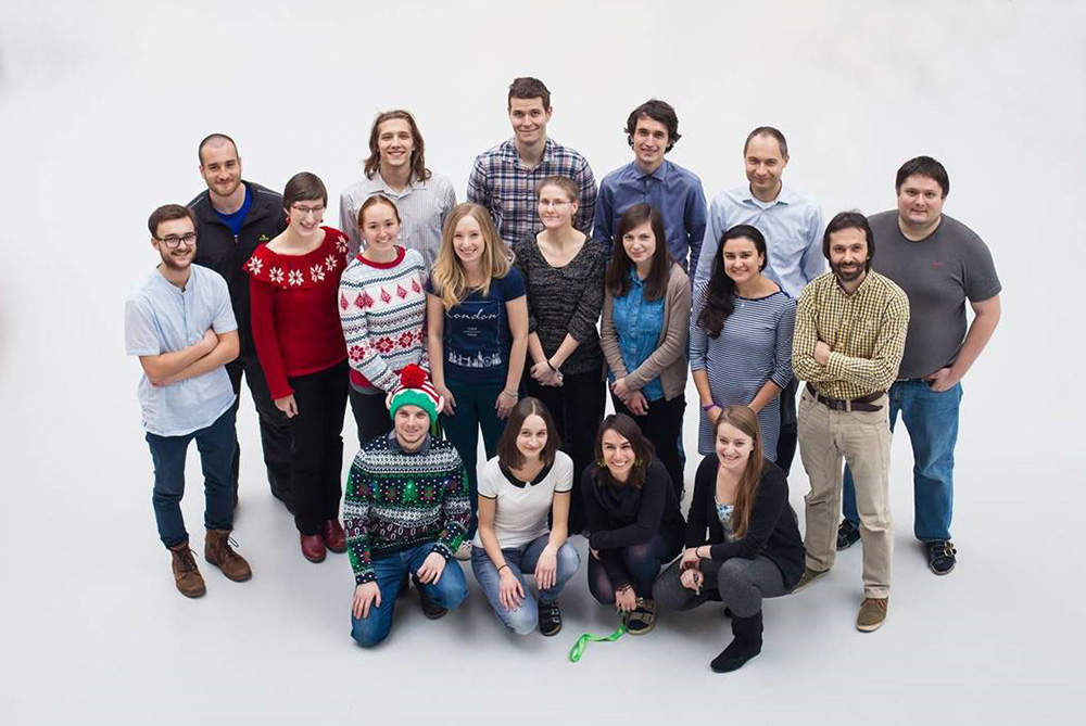
Research Group Leader

Postdoctoral Fellow

PhD student

PhD student

PhD student

PhD student

Laboratory technician

odborná pracovnice

Technician

odborná pracovnice ve výzkumu - postdoc

odborná pracovnice ve výzkumu - postdoc

Researcher

odborný pracovník - PhD student

odborný pracovník

odborný pracovník - PhD student

technička

odborná pracovnice ve výzkumu - postdoc

Researcher
1. Cryo-electron microscopy analysis of influenza A replication in vivo
Supervisor: Mgr. Pavel Plevka, Ph.D.
Annotation:
Influenza A virus causes hundreds of millions of infections, thousands of deaths, and economic losses of billions of US dollars every year. Still, the replication cycle of influenza A is not structurally characterized at molecular level. Cryo-electron microscopy allows observation of biological specimens in near-native state, but it faces the problem of decreased image quality for samples thicker than 300 nm. Focused ion beam milling can be used to prepare thin sections of vitrified cells to circumvent this issue. Subsequent cryo-electron tomography provides high-resolution structural information of complex cellular samples. Student will study replication and assembly of influenza A in vivo. Although three-dimensional structures of virus replication in vivo have been previously determined in fixed and stained cells, we intend on collecting data on cryo-preserved samples to enable the characterization of virus replication in near-native state without artifacts caused by heavy ion staining. The resulting tomograms will be analyzed with the help of pattern recognition and sub-tomogram averaging to obtain high-resolution details of the virus replication and assembly intermediates.
Keywords: Influenza A virus, replication cycle, cryo-electron microscopy, cryo-electron tomography


